
Grossology
Exploding caterpillars, cadaver labs, flesh-eating beetles, fish tongue parasites, dissected shark eyes – when it comes to gross science befitting Halloween, LSU Science has it all.
Check out our Top 5 Picks for gross scientific research experiences and discoveries in the LSU College of Science!
Gross Science #1: Cannibalistic Caterpillars, Exploding Caterpillars
In Bret Elderd’s lab in the LSU Department of Biological Sciences, it’s not uncommon to see caterpillar guts in little plastic cups and to witness caterpillars eating each other, for science. Bret, an associate professor of Biological Sciences, investigates how various factors – including cannibalism – affect disease transmission in insects, particularly in Lepidoptera, an order of insects including butterflies and moths.

Field-based experiment of disease spread in a population of insects inhabiting an individual plant. Credit: Bret Elderd.
Elderd was conducting field-based experiments with fall armyworm caterpillars (Spodoptera frugiperda) to mimic and study the spread of a lethal baculovirus in this lepidopteran when he noticed something strange. The experiments involved contaminating enclosed plants in the field with a lethal virus that eats caterpillars from the inside out, turning those who are infected into essentially swollen walking jelly bowls full of virus. Eventually the infected caterpillar splits open and oozes virus all over whatever it is eating – leading to infection of other caterpillars eating from the same plant. Gross. But each time Elderd went back into the field a few days after starting an experiment to collect his plants and fall armyworm samples, he would return with bags full of half-eaten fall armyworms.
“With the fall armyworms, we were losing a large portion of a number of samples we transported back to the lab because when the larvae got close to each other during transport, they started to eat one another,” Elderd said. “I started cursing my luck for conducting these studies with these dang bugs that would eat each other.”

A cannibalistic armyworm. Credit: Jose Bernardo Navarrete
It turns out that fall armyworm communities that cannibalize their virus-infected plant-mates have a better chance of surviving or even preventing a disease outbreak. Cannibalism may be gross, but it may be a smart move when a lethal disease is coming your way.
“What we show in a paper we published in American Naturalist is that if these caterpillars become cannibalistic and consume smaller, sick individuals in the population, transmission of this virus through the population is reduced,” Elderd said.

Bret Elderd holding a caterpillar in his lab. Photo by Paige Jarreau.
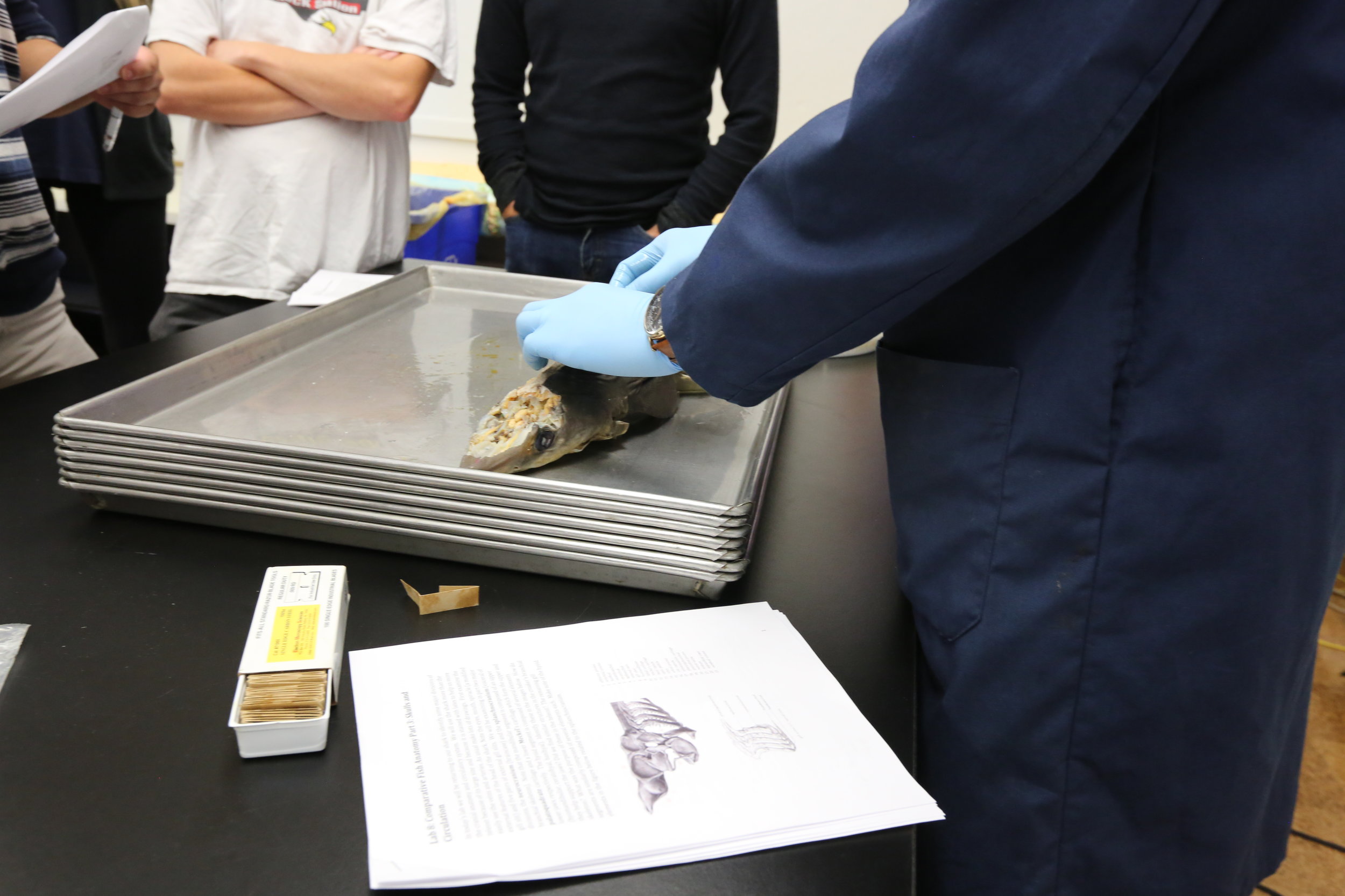
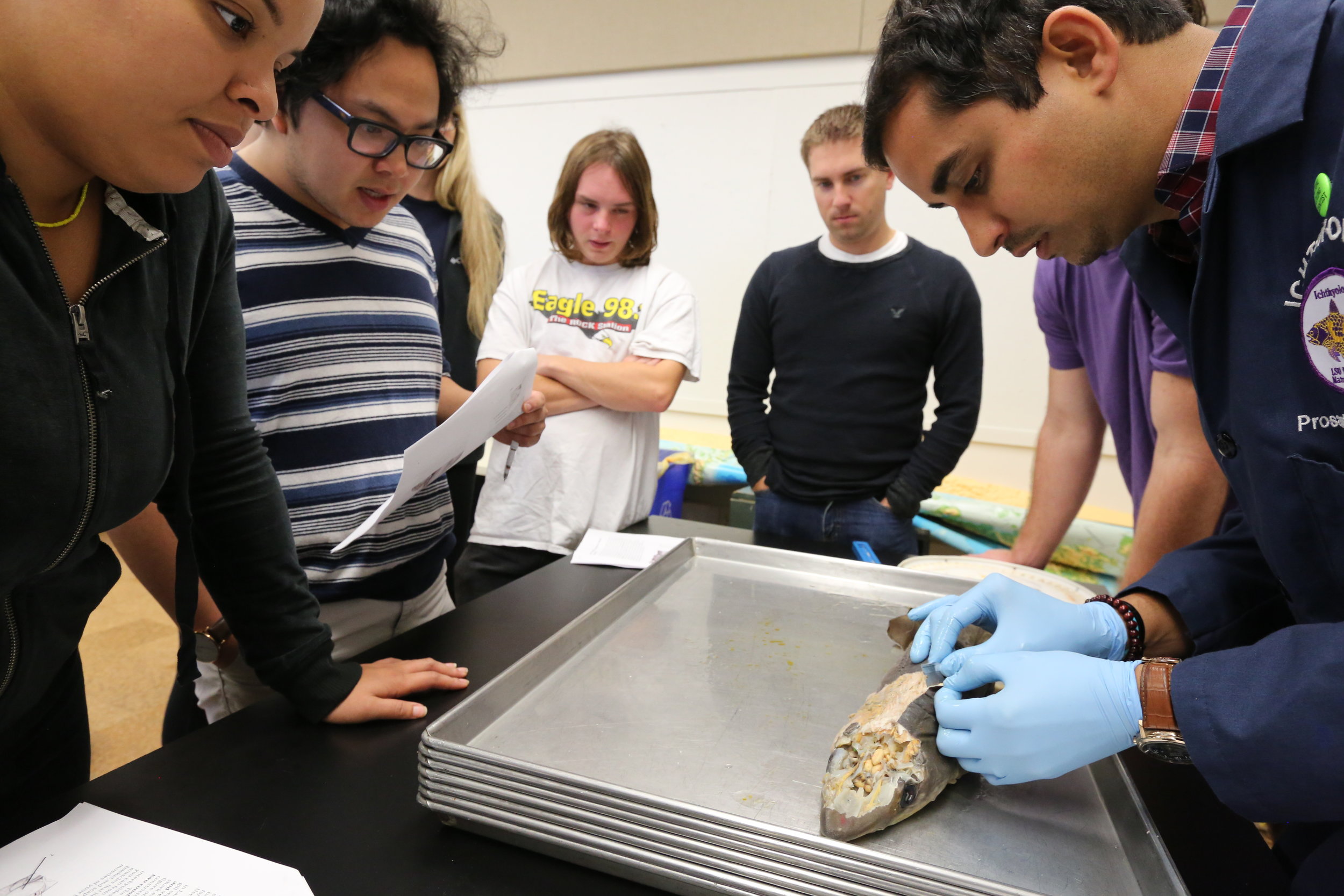
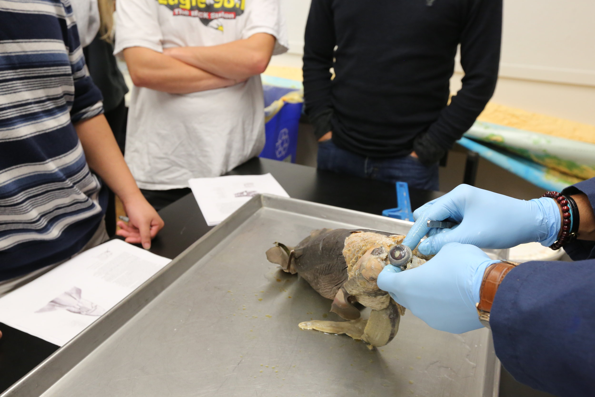
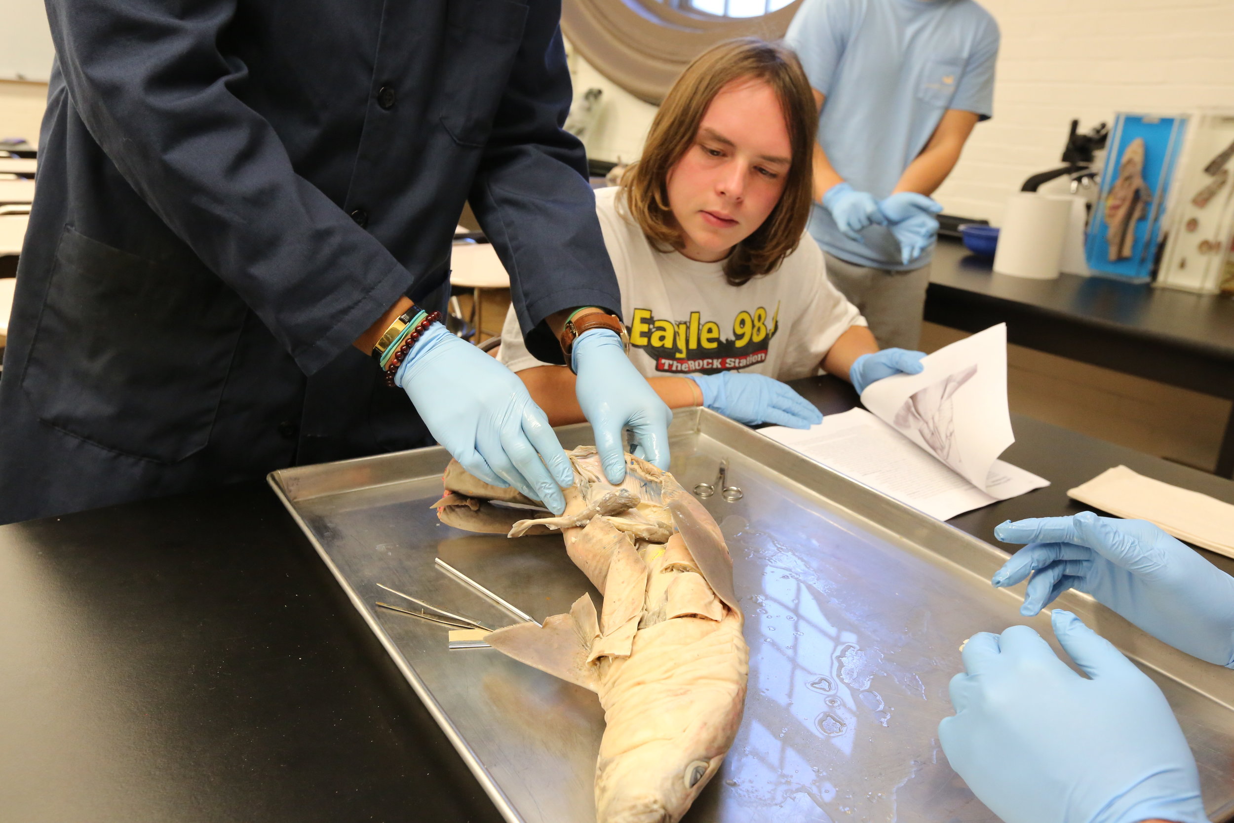
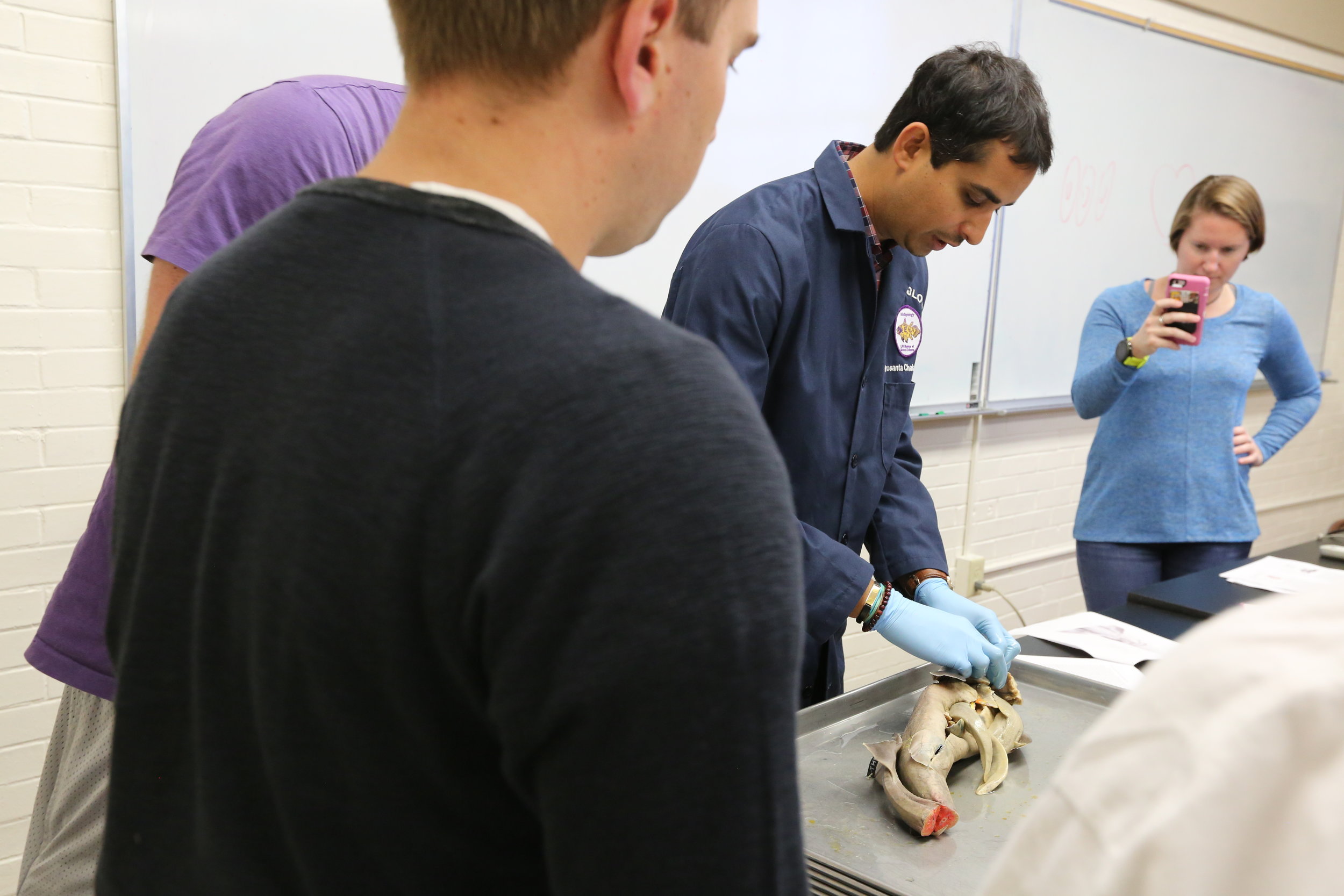
Gross Science #2: Dissecting Shark Eyes

Shark's Eyeball! Prosanta Chakrabarty demonstrates dissecting a shark in Biology 4145. Photo by Paige Jarreau.
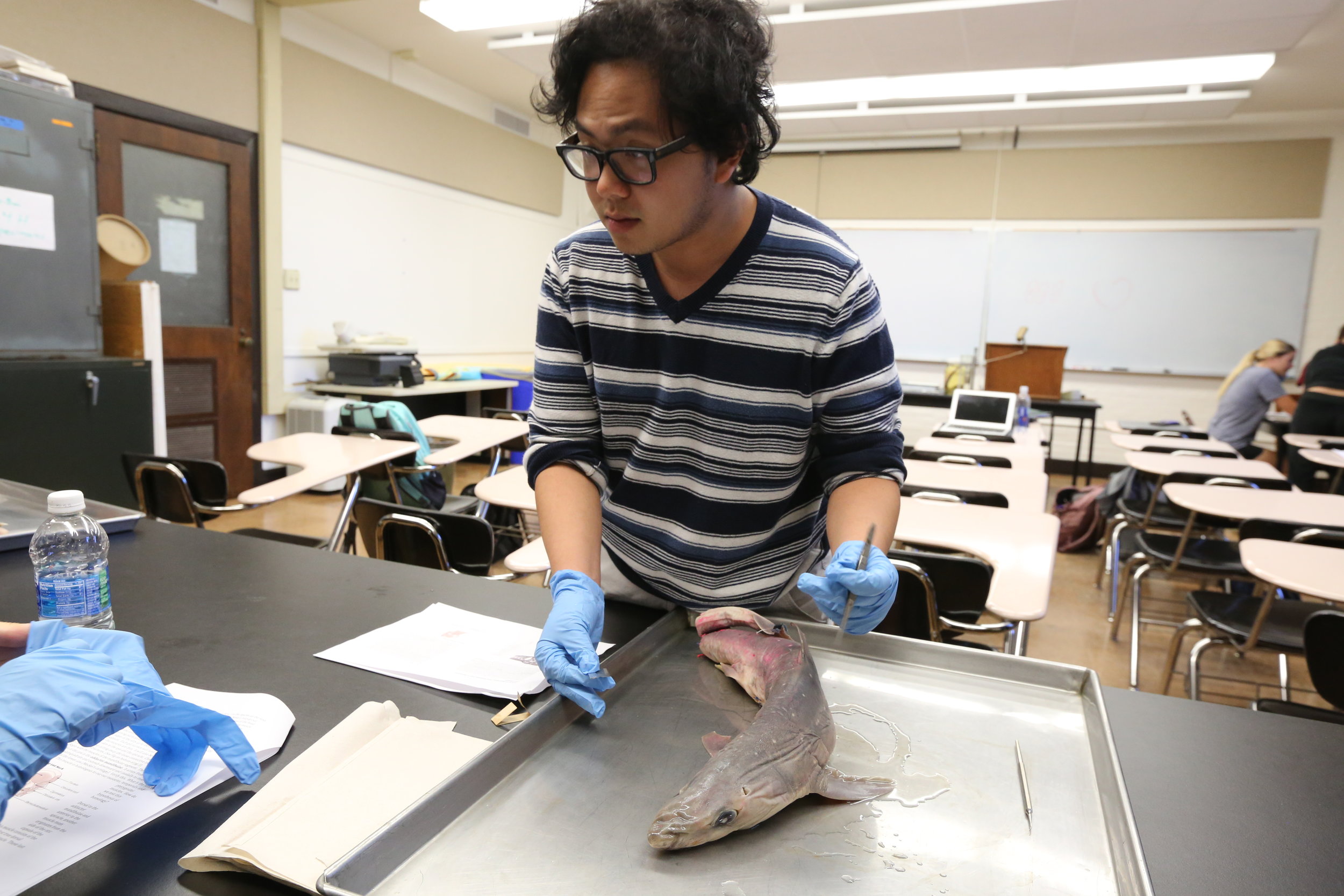
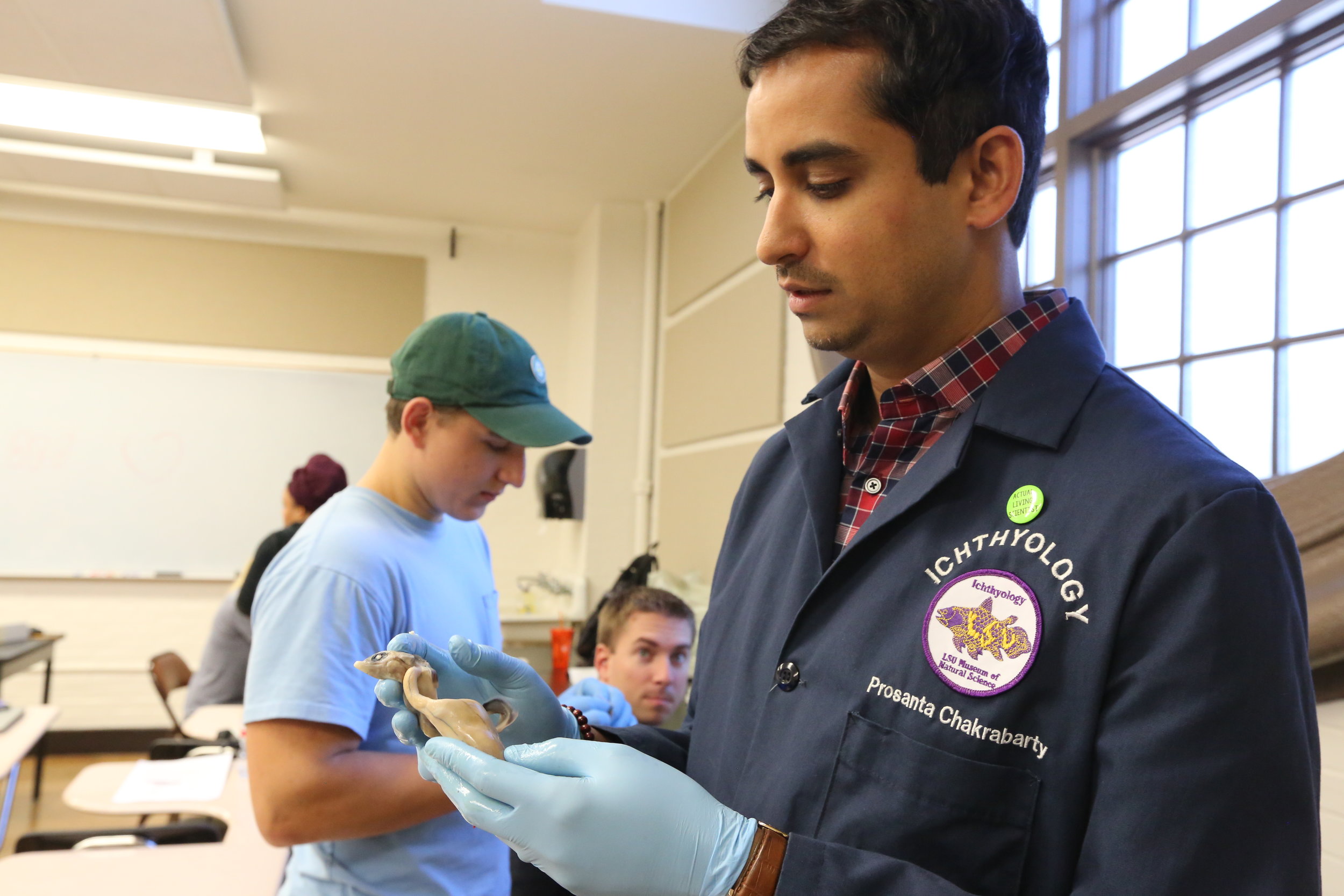
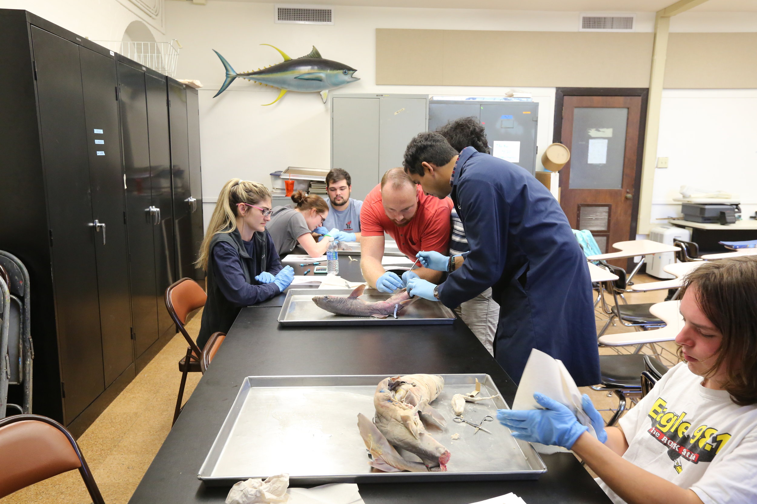
If cannibalistic and exploding caterpillars aren’t gross enough for you, you’d love Prosanta Chakrabarty’s ichthyology class Biology 4145, an upper division class on the biology and evolution of fishes. Just image having to carefully cut into a fluid-filled shark eyeball, opening it up and finding a round shiny lens, like a pearl inside of an oyster.
This semester, students in Prosanta’s class dissected dogfish sharks, revealing their brains and the inside of their eyes! Students dove into the cartilage of the head, examined the brain and nerves, and cut through the viscera following the flow of blood from the heart to the rest of the body. Through these dissections, students learned more about sharks’ cranial anatomy and circulatory system, and how these systems have homologues in humans. Muscles that raise the gills and work the jaws in these sharks are the same muscles that in humans now exist on our backs and even in our inner ears!
The dogfish sharks were farm-raised and injected with latex and dyes to help students visualize their veins and arteries. Prosanta’s students have also dissected a bony fish and reconstructed the bones of a boiled tilapia fish. But by far the coolest aspect of fish dissection, Prosanta says, is cutting open a shark’s eye to reveal its shiny marble-like lens.
“Except for the shape of the lens, much of the shark’s eye anatomy is just like ours,” Prosanta said.
Awesomely Gross.

Prosanta Chakrabarty demonstrates a dogfish shark dissection in his class at LSU. Photo by Paige Jarreau.
“In my class, students learn everything from the diversity of the more than 40,000 species of fishes, but also why it is important to learn the anatomy of these aquatic animals,” Prosanta said. “All vertebrates (including us) have aquatic ancestors, so most of our skeletal anatomy, organ systems and neural anatomy evolved originally to be adapted to the aquatic environment. In our dissection lab, we removed the skin around the head of a shark to show some of the muscles that are involved with lifting and moving the different elements of the cartilaginous jaws of these animals. We also discussed how these cartilaginous elements evolved into bony elements in terrestrial vertebrates that lack gills, like us. We did not evolve from sharks, but sharks have wonderfully preserved much of the early anatomy of the first jawed vertebrates.”


Gross Science #3: Cadaver Lab
Human cadaver labs are either gross or awesome, depending on your perspective. But they are valuable resources in training the next generation of medical professionals and researchers at LSU.

Arteries in the face and skull. Image via University of Liverpool Faculty of Health & Life Sciences, Flickr.com
“The opportunity to dissect a human body, or do hands-on study of a prosected body, is a rare opportunity and an incredible experience,” said Dennis Landin, William Prescott Foster Professor in the LSU School of Kinesiology. “Even after all these years, I’m still am amazed at the wonder of the human body! To hold a human heart in one’s hands, to see the inside of its’ chambers and the valves is very special. We remove the brains from each cadaver and that is also incredible. The students can directly see the cerebral circle, which they learn about in our neuroanatomy class, but there is no comparison to see the real thing. In addition to viewing normal human structures we often see anomalies and the results of surgical repairs. Our students see where blood vessels are positioned in coronary by-pass procedures, where pacemakers are inserted, mitral valve replacements, and hip and knee replacements, just to name a few.”
The School of Kinesiology at LSU is one very few programs nationwide, outside of medical schools, to offer human cadaver classes, and is the only program in the SEC to do so. Classes are held in the VetMed gross lab. Historically, enrollment in the course has been limited to Kinesiology majors. However, LSU College of Science Dean Cynthia Peterson arranged for space for a limited number of College of Science students in the lab beginning in summer 2017.
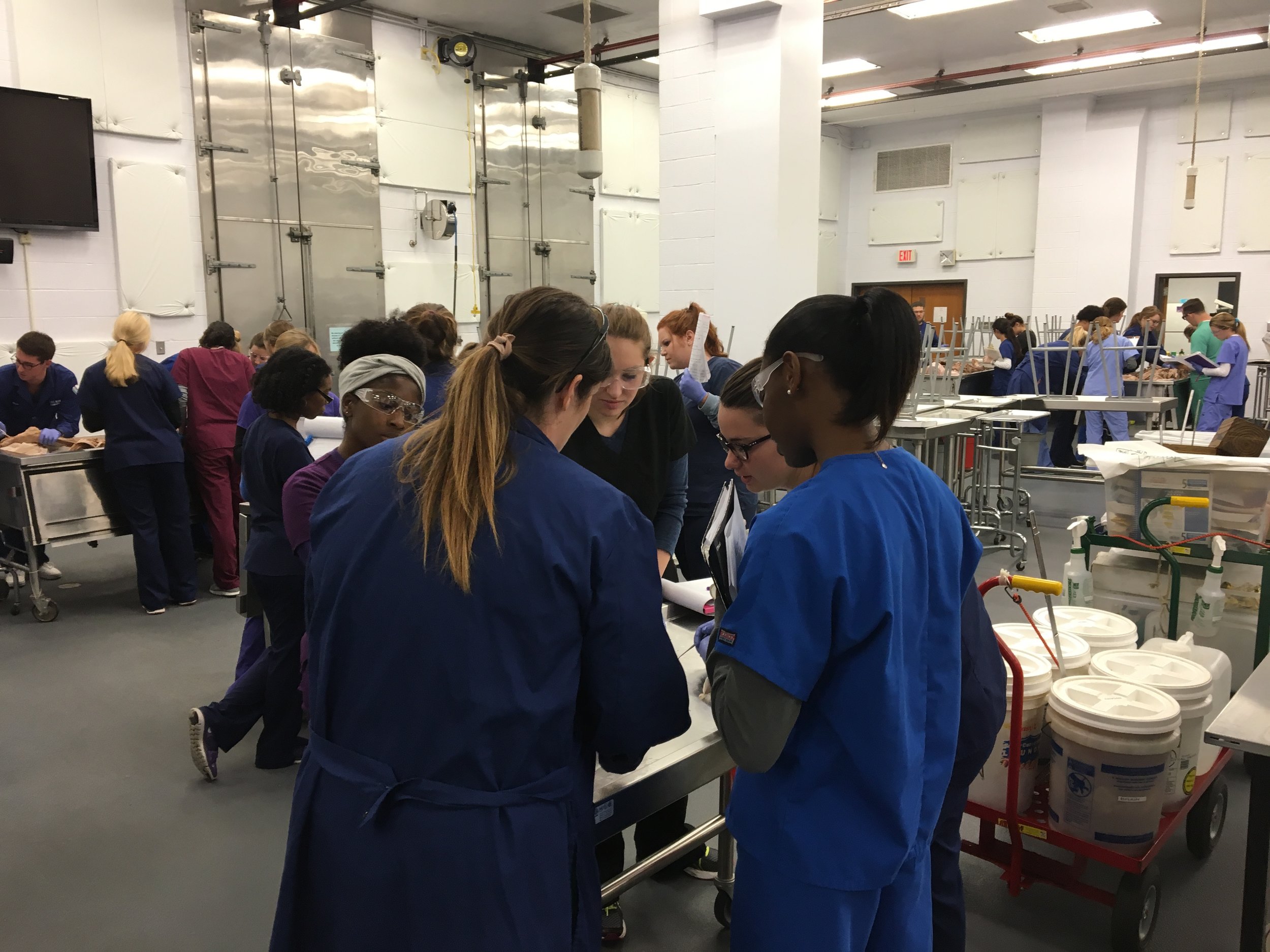
LSU students in the Cadaver Lab. Photo compliments of Dennis Landin.
Cadaver lab teaching assistant Haley Englade says she has several interesting stories from taking the lab herself.
“I had trouble at first staying in classroom the full hour because it made me a little nauseated,” Haley said. “The smell of formaldehyde is strong on particular days, and I would have to sit in the hallway with a wet towel on my neck. As a TA, I now have no problem in the class, as long as I eat a good breakfast."
"Students on the first day of class are often stunned at what they are seeing," Haley said. "It’s hard to imagine what the class will exactly be like. Because the lab is held in the LSU Vet School building, we often see dead animals being dissected on tables in the classroom. Our classroom is not a traditional teaching-style classroom; it contains big metal tables and cadaver tanks during class. This summer during the dissection class, my partner and I were working on the thorax and just as Dr. Landin reminded us about lab safety, I got some ‘juices’ in my eye. This is a big deal in the cadaver lab, because it could result in an infection. Dr. Landin and a few teaching assistants had to hold me up above the big sink in the classroom and pour an enormous amount of saline solution into my eye to flush out the possibilities of infection. That same day, another student at my tank got piece of flesh in his mouth, and all of us at the tank found it hysterical.”
Lawrence Messina, a biological sciences major who is in the Cadaver Lab this semester, enrolled in the course to gain advanced hands-on experience in preparation for medical school.
“There is nothing gross or creepy about the lab or the class itself,” Lawrence said. “The hardest thing to get used to is the smell of formaldehyde, but the cadavers are treated with respect as instruments of learning. Because of this course I have a much better grasp of the orientation of organs and the organization of nerves, blood vessels and muscle in the human body.”
Gross Science #4: A Fish’s Tongue Parasite

An isopod in a fish's mouth. Photo via Prosanta Chakrabarty.
What is grosser than looking into a fish’s mouth, and finding a bug instead of a tongue?!
LSU Museum of Natural Science Curator of Fishes Prosanta Chakrabarty was collecting a common croaker, Micropogonias undulatus, while fishing with other curators from the Museum in the Gulf of Mexico when he noticed something very strange about the fish.

An isopod in a fish's mouth. Photo via Prosanta Chakrabarty.
“My colleagues always joke that they only get weird stuff when they fish with an Ichthyologist,” Prosanta said. “This specimen was too small to keep, so I was about to throw it back in the water when I looked inside its mouth. Yuck!!! It had a tongue parasite!”
An isopod parasite had eaten the fish’s tongue and replaced that organ with itself. These parasites live with the fish they inhabit until the fish dies.
“It eats when the fish eats, getting little bits from whatever goes in the fish’s mouth,” Prosanta said. “I had known about these parasites, but this was the first time I ever saw one. Pretty gross!”
Gross Science #5: Flesh-Eating Beetles
Did you know that the LSU Museum of Natural Science has a flesh-eating bug room? The Dermestid beetle colony in the Museum may be gross to some, but it has a very important function in helping to prepare and preserve research specimens.
When Museum researchers collect animals in the field for research purposes, often including animals that have already been killed by cars and other accidents, they quickly begin the tedious process of preserving these specimens so that researchers who come after them can continue to learn more about our natural world and how to conserve it. After taking tissue samples and freezing them in liquid nitrogen for DNA analysis and other measurements, Museum researchers prepare specimens by skinning them and placing their skeletons in the LSU’s Bug Room, where the flesh-eating beetles get to work cleaning the bones. Researchers may also preserve soft tissues from specimens in jars of alcohol and formaldehyde for the Museum’s soft tissue collection.
Glaucia Del-Rio had to prepare many bird specimens from her recent to Brazil. We learn more from her on how specimens are prepared below!
Video: Glaucia Del-Rio prepping Elaenia mesoleuca
LSU College of Science: What is involved in preparing a bird specimen?
Glaucia: We make a complete dissection, taking data such as age, sex of the bird, breeding conditions, presence of parasites, stomach contents (what the bird was eating) and amount of fat (which indicates if the bird was in preparation for migration), among other data. We also collect pieces of muscle, heart, liver and intestines and freeze them in liquid nitrogen. The idea is to use these tissues to sequence DNA, RNA, proteins, or even to check for the presence of viruses and bacteria. We then clean and stuff the skin with cotton, which preserves the plumage patterns and colors. We write all the data on a small tag that gets attached to the scientific specimen.

A researcher pulls a frozen tissue sample from the liquid nitrogen tank at the LSU Museum of Natural Science. Photo by Paige Jarreau.
LSU College of Science: Why is specimen preparation necessary and important?

Photo of a Carolina parakeet specimen in the Museum of Natural Science. Photo by Paige Jarreau.
Glaucia: These specimens will be in our museum collections for hundreds of years, where they will be useful for describing new species. Many of the observations that lead Charles Darwin and Alfred R. Wallace to come up with ideas like the theory of evolution and natural selection were based on specimens that they had collected in the field, specially birds and insects.
In evolutionary biology, many discoveries and great ideas arise from simple looks at drawers full of stuffed bird skins collected from a wide range of geographies and time periods. Ecologists can measure museum bird specimens’ bills and wings to understand patterns of feeding behavior and seed dispersion. Breeding biologists can analyze breeding information from specimen tags to understand the breeding success of endangered birds across decades.
LSU College of Science: How did you learn to do this?
Glaucia: I learned from the best!!! Donna Dittman and Steve Cardiff, Collections Managers at the LSU Museum of Natural Science, first taught me how to prepare birds. During field trips in Brazil, Donna and Greg Schmitt, who are both great skinners, also gave me valuable tips to preparing bird skins.

Bird specimens at the LSU Museum of Natural Science. Photo by Paige Jarreau.
LSU College of Science: Is it gross?
Glaucia: I don't think that preparing birds is gross... although when we prepare vultures, the process can become very smelly! Overall, preparing scientific specimens is such an important task that I don't care to get my hands "dirty." When I prepare a specimen, I am not working only for me, or for my dissertation – I am leaving a legacy for the next generation of biologists. They will use these specimens to answer questions I have no idea can be asked... and that is really cool!